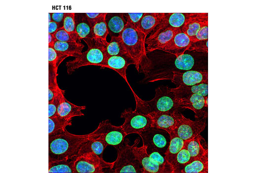
Actin filaments were labeled by dylight 554 phalloidin 13054 cell signaling technologies danvers ma usa.
Cell signaling anti lamin.
Antibodies used were polyclonal goat anti lamin b 6216 santa cruz biotechnology dallas tx usa.
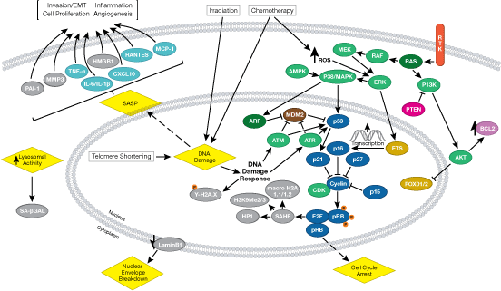
Lamin a c is cleaved by caspase 6 and serves as a marker for caspase 6 activation.
Type b lamins consist of lamin b1 and b2 encoded by separate genes 6 8.
During apoptosis lamin a c is specifically cleaved into a large 41 50 kda and a small 28 kda fragment 3 4.
Find out why customers rank cst highest for antibody specificity and sensitivity.
The cleavage of lamins results in nuclear dysregulation and cell death 5 6.
Lamin a and c are cleaved by caspases into large 41 50 kda and small 28 kda fragments which can be used as markers for apoptosis 4 5.
Wash three times for 5 min each with 15 ml of tbst.
An antibody shouldn t be one of the variables in your experiment.
Anti lamin a c antibodies are available from several suppliers.
During apoptosis lamin a c is specifically cleaved into a large 41 50 kda and a small 28 kda fragment 3 4.
The expected protein mass is 74 1 kda but there are 6 reported isoforms.
Incubate membrane with anti rabbit igg hrp linked antibody 7074 at 1 2000 and anti biotin hrp linked antibody 7075 at 1 1000 1 3000 to detect biotinylated protein markers in 10 ml of blocking buffer with gentle agitation for 1 hr at room temperature.
The cleavage of lamins results in nuclear dysregulation and cell death 5 6.
Lamin b1 is also cleaved by caspases during apoptosis 9.
In humans this protein is encoded by the gene lmna.
The protein may also be known as lamin c dhe lamin a cdcd1 cddc and cmd1a.
The cleavage of lamins results in nuclear dysregulation and cell death 5 6.
Lamin a c is cleaved by caspase 6 and serves as a marker for caspase 6 activation.
Phosphorylation of lamin a c at ser22 was identified in vivo in several cell lines by mass spectrometry analysis in proteomic screens.
Lamin a and c are cleaved by caspases into large 41 50 kda and small 28 kda fragments which can be used as markers for apoptosis 4 5.
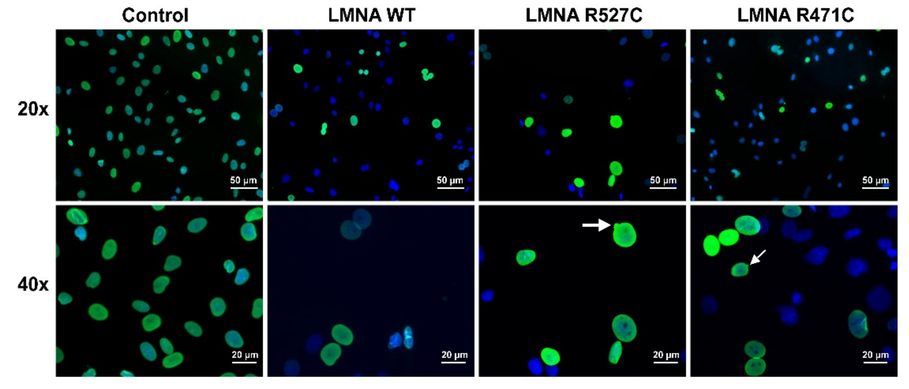
The cells were fixed in 2 paraformaldehyde for 10 min at room temperature.
2000 j struct biol 129 313 23.
Anti lamin a antibodies are readily available from several suppliers.
2000 j struct biol 129 313 23.
The full protein is reported to be 664 amino acid residues in length.
Proceed with detection section d.
Type b lamins consist of lamin b1 and b2 encoded by separate genes 6 8.
Lamin a is a reported synonym of the human protein lamin a c encoded by the gene lmna.